Single scan
In the Single scan tab you have:
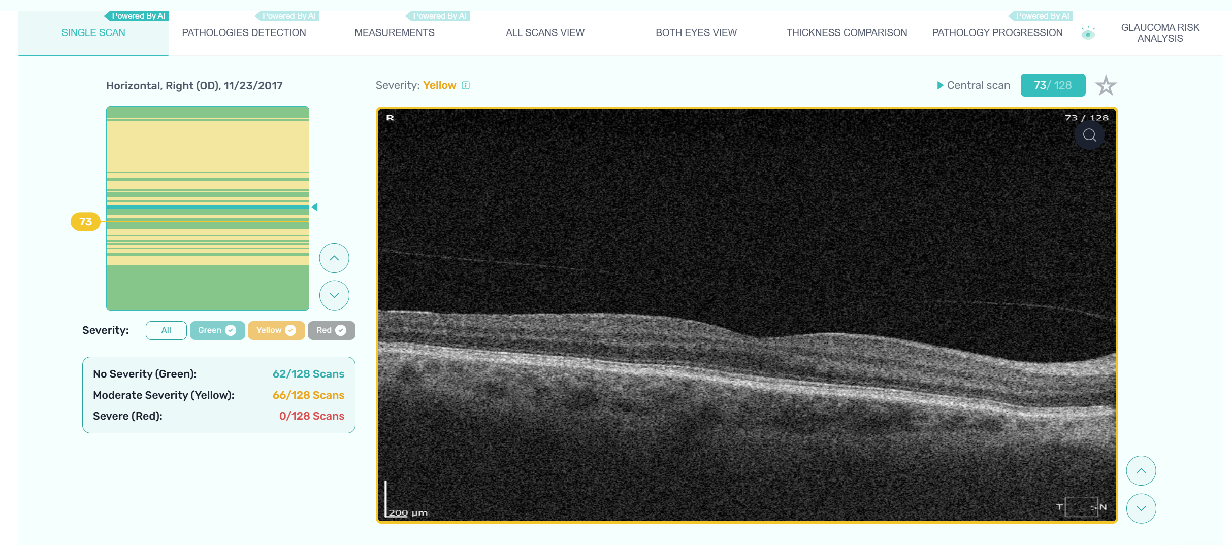
- Pathology severity visualization
When you upload N scans into the system, it will instantly differentiate between normal retina scans, moderate severity scans, and severe scans based on the degree of pathology found
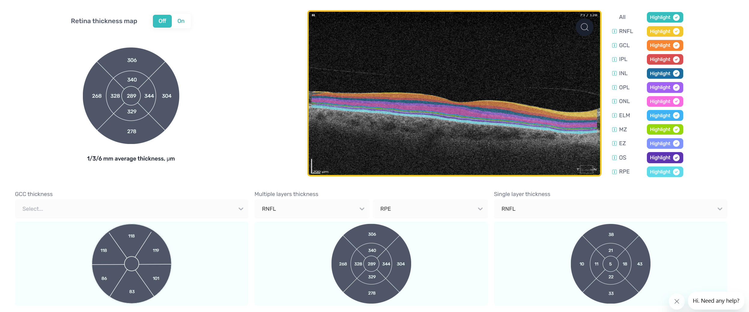
- Vendor neutral retina layers segmentation with AI
Altris AI can segment 10 retina layers and provide the possibility to explore each one of them
- Retina thickness, layers thickness, multiple layers thickness
Altris AI analyzes the thickness of the retina, separates layers or multiple layers
Single scan tab consists of:
-
OCT severity detection map with an option to browse through scans and select a specific one
-
Filter scans by severity order: all, green - normal retina, yellow - moderate concern, red - high concern
-
Arrows to change scan selection
-
Central scan
-
Scan number
-
Selected scan
-
Favorites (scans)
-
Scan zoom icon (user can review a particular scan in its original size)
-
Layer thickness (average, single, multiple, GCC thickness)
-
Heatmap (ON/OFF)
-
Segmentation and separation of 10 retinal layers with an option to select single or multiple layers
* All names included in this User Manual are not real and are for demonstration purposes only
Once the scan has finished uploading, the system classifies the scans by severity and detects pathological scans that require further analysis immediately. All scans are colored by their severity: green - normal retina, yellow – moderate with slight deviations, and red – severe, which means there are significant changes in retina layers.

You can filter scans to see only particular severity groups.
You can easily navigate through the scans with arrow buttons.
Altris AI detects low-quality scans automatically and warns you about the possibility of poor-quality data which will affect the quality of the segmentation and classification.
Altris AI segments 10 retina layers and provides the possibility to explore each one of them separately - NFL, GCL, IPL, INL, OPL, ONL, ELM, MZ, EZ, RPE, Choroid.
NFL – Nerve Fiber Layer; GCL – Ganglion Cell Layer; IPL – Inner Plexiform Layer; INL – Inner Nuclear Layer; OPL – Outer Plexiform Layer (synaptic); ONL – Outer Nuclear Layer; ELM – External Limiting Membrane; MZ - Myoid zone of the photoreceptors; EZ – Ellipsoid Zone(AKA IS/OS Junction); RPE –Retinal Pigment Epithelium.


Altris AI analyzes the thickness of retinal layers, separate layers, or multiple layers.
Altris AI uses the following parameters to calculate layer sickness:
- Axial - less precise, used in OCT
- Minimum distance - more precise, more time-consuming to calculate
To present these measurements 4 ETDRS circles are introduced on the platform:
- The total average thickness of the layers
From RNFL till RPE

- Single layer thickness
ETDRS calculation for only one of the selected layers is shown
RNFL, GCL, IPL, INL, OPL, ONL, ELM, MZ, EZ, RPE

- Multiple layers thickness
From: RNFL, GCL, IPL, INL, OPL, ONL, ELM, MZ, EZ
To: GCL, IPL, INL, OPL, ONL, ELM, MZ, EZ, RPE

- GCC thickness
The following two calculations are given:
GCC (GCL+IPL)
GCC + RNFL (RNFL+GCL+IPL)

Heatmap ON mode

Updated 24 days ago